All posts by Meagan

DVT 101

Looking for deep vein thrombosis (DVT) of the lower extremity is a common request. The most important aspect of a DVT ultrasound is the compression of the vein.

On ultrasound, vessels look black, or anechoic, due to the fact that they are fluid filled. You can also use colour Doppler to see the flow of blood within these vessels.

Holding your probe in transverse on the patient’s leg, find the common femoral artery and vein at the proximal thigh. By applying firm pressure with the probe to the patient, the vein should easily collapse. Follow the vein from the thigh to behind the knee, to the popliteal vein.

sfv-comp-norm.jpg

However, if the vein does not compress, this should raise suspicion for a DVT. Often, you will be able to see clot within the vein, as the vessel is no longer anechoic, but filled with hypoechoic clot. Also, if you turn on colour Doppler there will be no colour flow within the vessel.

DVT +

clot

Images:

http://www.nuemblog.com/dvt

https://radiopaedia.org/cases/lower-limb-deep-venous-thrombosis-dvt

Save

What’s in a name? Different types of ultrasound probes

Types of Probes:

There are three main types of ultrasound probes: curvilinear, linear and phased.

Curvilinear. This is a low frequency probe with a curved face. Typically used for abdominal, gyne, obstetrical and pleural scanning. The curved face gives and increased field of view, while the lower frequency allows for better penetration.

Linear: High frequency probe with a flat, rectangular face. Used for vascular, small parts and musculoskeletal scanning. Due to shape of probe, image is rectangular, not a curved arc. *Tip: Most units have an option to make the image widescreen, increasing the field of view.

Phased: Low Frequency rectangular shaped probe with a small face. Primarily used for cardiac scanning (echocardiography). The small face of the probe allows it to fit between ribs, but the arrangement of crystals creates a curvilinear field of view.

Aorta and IVC

Ultrasound can be used to assess the main vascular structures in the abdomen. The most common examination is to look for an Abdominal Aortic Aneurysm (AAA).

To find your aorta, start by placing the probe on the patients’ abdomen in transverse, just below the xiphoid process. You will see anechoic oval and circle, above a crescent shadow. These represent the IVC, Ao and Vertebra, respectively. Using firm pressure, slide probe downwards toward the umbilicus, at which you should see the Ao bifurcate into the Iliac arteries. Once found in transverse, try this motion while the probe is in the sagittal plane.

AO IVC TRaortanormal


An aortic aneurysm is a focal dilation of the Aorta, and should be greater than 3.0 cm when measured from anterior to posterior.

aaa

The IVC is located just right of midline, and can be seen by the liver. It is best appreciated in the sagittal plane. Notice how the IVC is phasic, changing size with respiration. If you ask your patient to breathe in, or “sniff”, you should see the vessel collapse more than 50%, demonstrating a normal venous pressure.

C60_Liver_IVC_Midline[3]_1

Images:

http://reference.medscape.com/features/slideshow/ultrasound

http://www.sonosite.cn/clinical-media


Save

Image Optimization

Depth, Focus, Gain

When it comes to image quality, if you follow these easy steps for every image, you can’t go wrong! First, adjust your depth. Is the structure you are imaging, say the aorta, way at the top of the screen? Decrease your depth so your image ends just below the area of interest. Is the lower pole of the kidney cut off? Increase your depth to include a few centimeters below the kidney border.

Now that you’ve adjusted your depth, apply the focus! By tapping or twisting the focus knob, place your focal zone at or just below the structure of interest. This will place optimal resolution at the organ you want to see.

Finally, tweak the gain. Usually a dial on the main console, twist to increase or decrease the brightness of the image. Your goal: Make anechoic structures, such as vessels or the bladder black, but still bright enough to see surrounding structures.

With these three steps, your images will be picture perfect!

What do Gallstones look like?

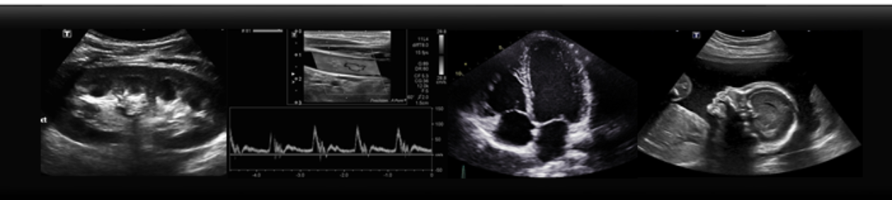
Cholecystitis is one of the more common ailments seen in emergency departments, and one of the most frequent indications for an abdominal ultrasound. Gallstones will appear as echogenic, or bright white, round stones within the gallbladder, with shadows posterior. The gallbladder itself will appear anechoic, or black, tubular structure, adjacent to the right lobe of the liver and the main portal vein. When assessing for cholecystitis here are some other sonographic signs:

gallstones-1acute-calculous-cholecystitis


-Look for mobility. Do the stones roll when you move the patient? Look at the gallbladder supine, stone will be sitting towards the “neck” of the gallbladder, because that is dependent location. Now try tolling your patient LLD, or even sitting up. The stones should roll to the fundus! If not, are they stuck in the neck?

acute-cholecystitis-with-gallbladder-neck-calculus

– Is the gallbladder wall thick? Measure the wall in transverse and it should be less than 3 mm

acute-acalculous-cholecystitis

-Is there free fluid? Look for a pointy, “triangle” shaped anechoic area beside the GB.

acute-calculous-cholecystitis-ff

– Is there a positive Murphy’s Sign? When you apply pressure to the gallbladder, does this correspond with the patient’s area of pain? Often the response is quite clear.


Images courtesy of Radiopedia.org:

Case courtesy of Dr Derek Smith, <a href=”https://radiopaedia.org/”>Radiopaedia.org</a>. From the case <a href=”https://radiopaedia.org/cases/42795″>rID: 42795</a>

Case courtesy of Dr Andrew Dixon, <a href=”https://radiopaedia.org/”>Radiopaedia.org</a>. From the case <a href=”https://radiopaedia.org/cases/9558″>rID: 9558</a>

Case courtesy of RMH Core Conditions, <a href=”https://radiopaedia.org/”>Radiopaedia.org</a>. From the case <a href=”https://radiopaedia.org/cases/38022″>rID: 38022</a>

Case courtesy of Dr M Osama Yonso, <a href=”https://radiopaedia.org/”>Radiopaedia.org</a>. From the case <a href=”https://radiopaedia.org/cases/16769″>rID: 16769</a>

Save

Save

The Gallbladder

It’s Thanksgiving weekend, and what a fitting time to talk about the gallbladder. With the rich meals associated with the holidays, it’s no surprise that the emergency department fills with patients who have biliary tract disorders such as gallstones and cholecystitis.

Here are some tips on how to find the gallbladder on ultrasound. Begin with the patient supine, placing probe sagittally and to the right of mid-line. The image should show liver and the gallbladder. Often a held inspiration can help move obscuring bowel gas. Now often patients do not have a nice supine window, so next view to try is intercostal. Place the probe at right lateral position, scanning between the ribs. Next look at the gallbladder when the patient is in a left lateral decubitus position. Often this is the best view to look at the GB because the liver creates an acoustic window and the bowel moves to the left. Again, place probe subcostally, right of mid-line, and sweep with your probe in the sagittal position.

Below is an ultrasound image of a normal gallbladder. Stay tuned for further posts about gallbladder pathology!

normal-upper-abdominal-ultrasound-male-adult

Case courtesy of Dr Henry Knipe, <a href=”https://radiopaedia.org/”>Radiopaedia.org</a>. From the case <a href=”https://radiopaedia.org/cases/48019″>rID: 48019</a>

Sonography Week 2016!

This week is Sonography Week, celebrating ultrasound and the dedication of medical professionals who use it. Below is a link to an interesting video demonstrating the development of ultrasound in healthcare. Hopefully it will inspire you to continue to learn about all that ultrasound can do!

Video Courtesy of Society of Diagnostic Medical Sonography.

https://www.youtube.com/watch?v=sVa7PJm2okk

Assessment is Key!

It cannot be stressed enough that the real shining star of ultrasound is the real-time assessment of a structure; the actual sweeping back and forth through the organ of interest. It can be all too easy for new scanners to focus on finding the organ and taking a picture of it. Don’t forget to sweep through that structure! Here’s a good example: When scanning a kidney in the sagittal plane, plant your probe and angle it side to side. As you sweep through the kidney, assess medial to lateral until you have scanned completely in and out of the kidney. You never know, you might pick up an exophytic cyst on the periphery of the kidney!

Don’t be discouraged if you aren’t getting textbook images at the beginning of your ultrasound journey, it’s the assessment that counts.

Key

Save