Tag Archives: ED

Aorta and IVC

Ultrasound can be used to assess the main vascular structures in the abdomen. The most common examination is to look for an Abdominal Aortic Aneurysm (AAA).

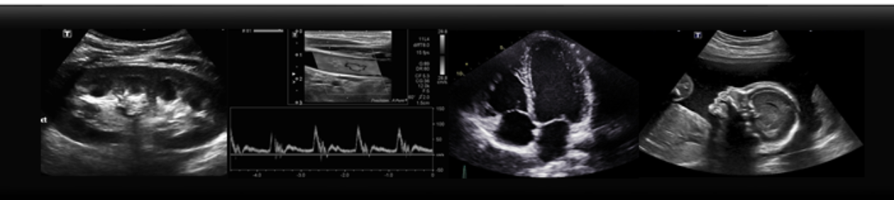
To find your aorta, start by placing the probe on the patients’ abdomen in transverse, just below the xiphoid process. You will see anechoic oval and circle, above a crescent shadow. These represent the IVC, Ao and Vertebra, respectively. Using firm pressure, slide probe downwards toward the umbilicus, at which you should see the Ao bifurcate into the Iliac arteries. Once found in transverse, try this motion while the probe is in the sagittal plane.

AO IVC TRaortanormal


An aortic aneurysm is a focal dilation of the Aorta, and should be greater than 3.0 cm when measured from anterior to posterior.

aaa

The IVC is located just right of midline, and can be seen by the liver. It is best appreciated in the sagittal plane. Notice how the IVC is phasic, changing size with respiration. If you ask your patient to breathe in, or “sniff”, you should see the vessel collapse more than 50%, demonstrating a normal venous pressure.

C60_Liver_IVC_Midline[3]_1

Images:

http://reference.medscape.com/features/slideshow/ultrasound

http://www.sonosite.cn/clinical-media


Save

What do Gallstones look like?

Cholecystitis is one of the more common ailments seen in emergency departments, and one of the most frequent indications for an abdominal ultrasound. Gallstones will appear as echogenic, or bright white, round stones within the gallbladder, with shadows posterior. The gallbladder itself will appear anechoic, or black, tubular structure, adjacent to the right lobe of the liver and the main portal vein. When assessing for cholecystitis here are some other sonographic signs:

gallstones-1acute-calculous-cholecystitis


-Look for mobility. Do the stones roll when you move the patient? Look at the gallbladder supine, stone will be sitting towards the “neck” of the gallbladder, because that is dependent location. Now try tolling your patient LLD, or even sitting up. The stones should roll to the fundus! If not, are they stuck in the neck?

acute-cholecystitis-with-gallbladder-neck-calculus

– Is the gallbladder wall thick? Measure the wall in transverse and it should be less than 3 mm

acute-acalculous-cholecystitis

-Is there free fluid? Look for a pointy, “triangle” shaped anechoic area beside the GB.

acute-calculous-cholecystitis-ff

– Is there a positive Murphy’s Sign? When you apply pressure to the gallbladder, does this correspond with the patient’s area of pain? Often the response is quite clear.


Images courtesy of Radiopedia.org:

Case courtesy of Dr Derek Smith, <a href=”https://radiopaedia.org/”>Radiopaedia.org</a>. From the case <a href=”https://radiopaedia.org/cases/42795″>rID: 42795</a>

Case courtesy of Dr Andrew Dixon, <a href=”https://radiopaedia.org/”>Radiopaedia.org</a>. From the case <a href=”https://radiopaedia.org/cases/9558″>rID: 9558</a>

Case courtesy of RMH Core Conditions, <a href=”https://radiopaedia.org/”>Radiopaedia.org</a>. From the case <a href=”https://radiopaedia.org/cases/38022″>rID: 38022</a>

Case courtesy of Dr M Osama Yonso, <a href=”https://radiopaedia.org/”>Radiopaedia.org</a>. From the case <a href=”https://radiopaedia.org/cases/16769″>rID: 16769</a>

Save

Save